IDEXX case study: Anaplasmosis in a dog
Clinical Pathology Case Study: Anaplasmosis in a dog
Background information
| Age: | 3 years old |
| Sex: | Male (neutered) |
| Species: | Canine |
| Breed: | Eskimo dog |
| History: | Two-day history of decreased appetite and lethargy |
Clinical signs
- On physical exam he is febrile.
- A complete blood count reveals a mild thrombocytopenia.
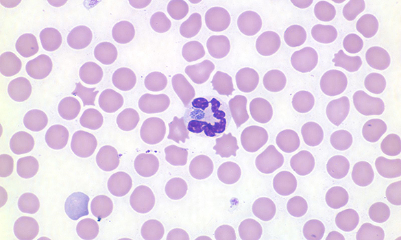
An image of the blood smear
Cytologic description and interpretation
Thrombocytopenia is confirmed with only 4-5 platelets per 100x field. In addition, several neutrophils contain aggregates of basophilic cocci bacteria, consistent with Anaplasma or Ehrlichia morula.
Interpretation: Based on the dog’s geographic location and travel history, Anaplasma phagocytophilum is the presumed organism.
Discussion
Anaplasmosis (typically Anaplasma phagocytophilum) is a tick-borne infection found worldwide in a variety of animals. Most infections are asymptomatic or cause short-lived, mild clinical signs (eg. anorexia, lethargy, stiffness, fever). Thrombocytopenia is the most common laboratory abnormality. The thrombocytopenia is typically mild and is not associated with bleeding abnormalities.
In dogs, Anaplasmosis has a similar presentation to other tick-borne diseases, including Lyme disease and Ehrlichiosis. Anaplasma and Ehrlichia organisms can occasionally be seen within leukocytes of acutely-infected animals. If found, the organism and patient’s geographic location can be used to reach a definitive diagnosis. However, visible organisms are only present in acute infections and sometimes in very low numbers. Therefore, PCR testing, which is more sensitive than blood smear evaluation, is another diagnostic option.
In areas where Anaplasmosis is common, many dogs are exposed subclinically to A. phagocytophilum and develop antibodies that can persist for 6 months after infection. A positive antibody test therefore should always be interpreted in combination with the patient’s clinical signs.
About the author
Julie Webb DVM, DACVP
Dr.Webb is a native of Ottawa and received her DVM from the Ontario Veterinary College in 2003. She practiced in the Ottawa area for 2 years before pursuing a residency in clinical pathology at the University of Georgia. She became a Diplomate of the American College of Veterinary Pathologists in 2008 and worked as a clinical instructor at the University of Wisconsin-Madison from 2008 to 2015: performing diagnostic service, training residents and teaching veterinary students. She joined IDEXX in February of 2015.
In her spare time, Dr. Webb enjoys spending time with her dogs and staying active by cycling, swimming, and playing hockey and soccer. She also enjoys gardening in the summer as well as working with stained glass during the winter months.
Should you have any questions about this case or wish to discuss the diagnosis in greater detail, please do not hesitate to contact the author.
View clinical pathology case studies
NEW: Dermatophytosis in a dog, by Caroline Piché, DMV, IPSAV, MSc, Diplomate ACVP
Anaplasmosis in a dog, by Julie Webb, DVM, DACVP
Blastomycosis in a dog, by Julie Webb, DVM, DACVP
Cryptococcus in a dog, by Carrie Flint, DVM, DACVP
Cutaneous mycobacteriosis (Leprosy) in a cat, by Natalie Kowalewich, DVM, DACVP
Demodex in a dog, by Brittney Fierro, DVM, DACVP
Feline infectious peritonitis in a cat, by Heidi Peta DVM, MVSc, DACVP
Mammary fibroepithelial hyperplasia in a young cat, by Emmeline Tan, DVM, DVSc, DACVP
Microvascular dysplasia in a dog, by Cathy Monteith DVM, MVSc, DACVP
Severe hyperglobulinemia in a dog, by Sébastien Overvelde, DVM, MSc, DACVP
Reference:
Greig B and Armstrong PJ. Canine Granulocytotropic Anaplasmosis. In: Infectious Disease of the Dog and Cat, 3rd ed., 2006, p. 219-224.
All case studies were prompted by real submissions to IDEXX Canada pathologists at one of our reference laboratories.
To protect the confidentiality of our customer and client, the background information has been slightly modified.