IDEXX inVue Dx Cellular Analyzer
Fine needle aspirate cytology
Test more patient samples. Take more informed next steps than ever before.
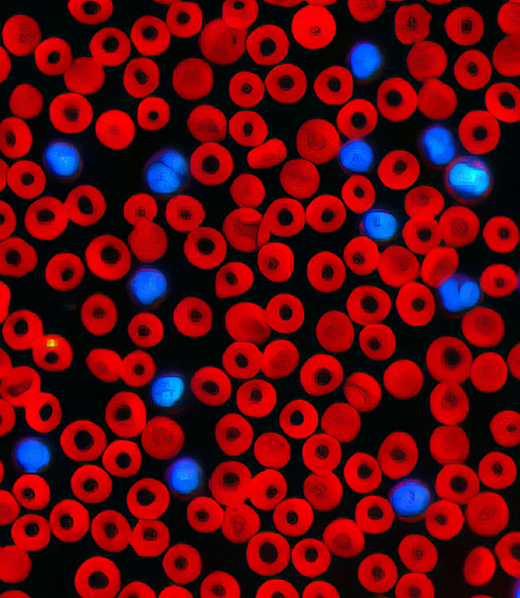
The IDEXX inVue Dx Cellular Analyzer identifies, measures, and quantifies cells to provide objective information that supports your diagnostic plan.*
- Get identification of a nonviable sample quickly.
- Receive results and next-step guidance for common skin masses during the patient visit.
- If you need an expert review, send your results to a clinical pathologist at IDEXX Reference Laboratories, no slides required.
- Stay ready for an expanding menu in future.





How practices are confidently putting objectivity to practice.
When veterinary teams talk about what’s working in their clinics, the results speak for themselves. Take a look at what veterinarians and their teams are saying about the IDEXX inVue Dx analyzer.
“We are sampling more growths than ever before. Previously we had to rely on what we saw under the microscope and decide if we should send it off for cytology...Now we are getting real-time results and able to make decisions quickly about moving forward with surgery sooner.”
—Elizabeth Nguyen, DVM
Preparing samples
The best results begins with the best sample. It’s the foundation of every FNA. The three steps below can help improve sample quality and diagnostic success with FNA.
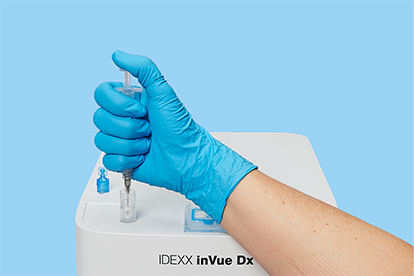
1. Expel the aspirate into the sample tube.
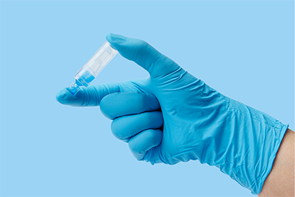
2. Cap the tube and invert it 5 times to mix.
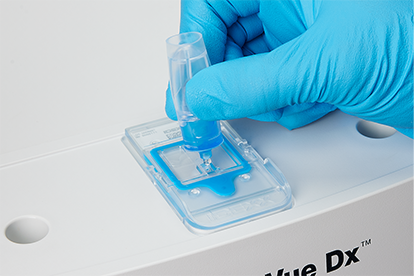
3. Remove the tab and pour the entire tube contents into the cartridge port.
Here's a short video to walk you through how to prepare an FNA sample for analysis.
Understanding results
Only you can evaluate your patient’s clinical signs. Add trusted diagnostics and clear, shareable images from the IDEXX inVue Dx Cellular Analyzer, and you have the information you need to make informed clinical decisions and help pet owners better understand your recommendations.
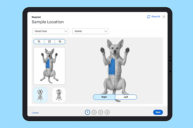
Diagnostic insights with structured inputs
Adding sample and lesion details on the IDEXX VetLab Station helps you capture all of the pertinent case information, save it to the patient record, and share it when a pathology review is requested.
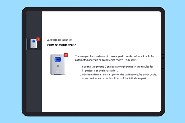
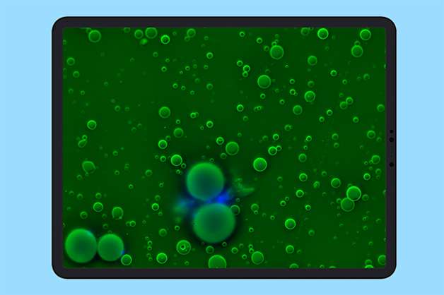
Sample quality assessment
The analyzer evaluates whether the sample contains sufficient diagnostic material for interpretation.
Common lesion classification
Mast Cell Tumor, adipose tissue and keratinized growth results are reported as suspected or detected based on the cellular characteristics identified in the sample.
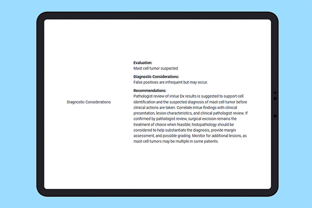
Diagnostic considerations
Result classifications help support clinical decision-making and guide appropriate next steps, including additional testing or treatment.
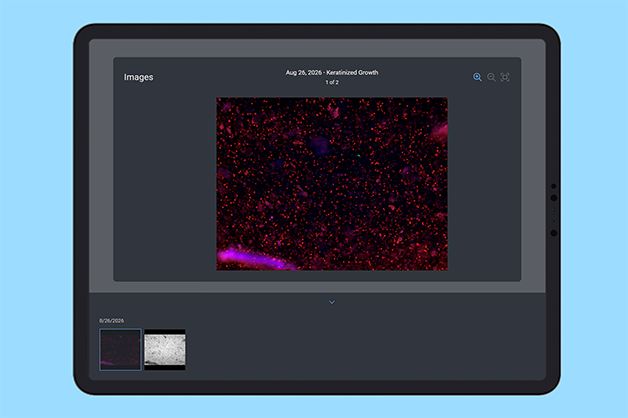
Image gallery
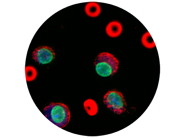
To visually support client conversations, the image gallery pairs analyzer results with clear visuals similar to reference lab pathology images.
Take the subjectivity out of interpretation.
With certainty comes confidence. The IDEXX inVue Dx Cellular Analyzer's reporting structure takes the guesswork out of interpreting cytology results, bringing clear, objective language to FNA sample evaluation, right in your clinic, in minutes. For general practitioners navigating real-world caseloads, that means consistent, defensible answers you can act on with confidence.
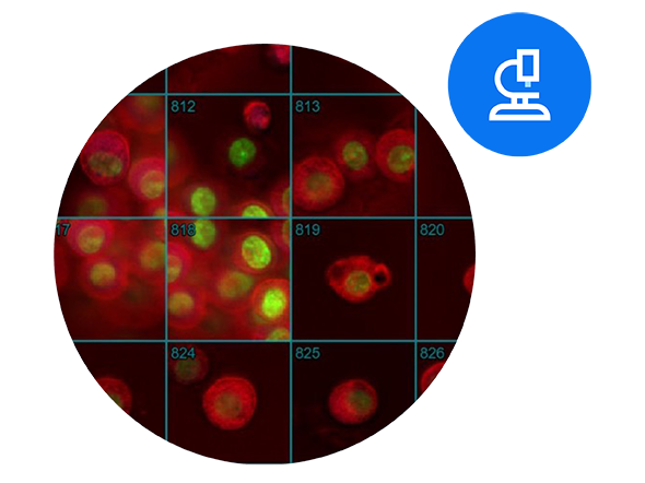
Clinical pathology evaluation: Expert support when you need it.
In many cases, the automated findings are enough to guide the next clinical step. When further insight and evaluation are needed, you can send your IDEXX inVue Dx FNA results and images as well as your case details to an IDEXX Reference Laboratories pathologist for review.
The Curated Collection: IDEXX inVue Dx Cellular Analyzer
Thoughtfully organized, carefully chosen education resources for veterinary professionals.
Frequently asked questions
- Initiate the sample run on the IDEXX VetLab Station (be sure to select IDEXX inVue Dx and FNA when prompted).
- Prepare the sample (see “Preparing samples” above).
- Insert the prepared cartridge into the IDEXX inVue Dx analyzer.
- Press the Start button on the analyzer.
- Slide-free workflow and affordability increases access to cytology for patients with lumps and bumps.
- Pathologist-trained AI provides consistent, objective results with reference laboratory-level accuracy on reported parameters.
- Results, images, and diagnostic considerations are available quickly in-house, allowing veterinarians to discuss next steps with pet owners during their visit.
- Results will be available in approximately 15 minutes; however, it could take less or more time depending on sample conditions.
- If the sample is nonviable (not enough or poor-quality cells), notification is provided in approximately 5 minutes or less.
- Validated: Canine cutaneous and subcutaneous masses
- Not supported: Lymph node, fluid, or organ samples
Resources
Just getting started with the IDEXX inVue Dx analyzer? We have a selection of guides, videos, and other resources that make it easy to get up to speed.
Test menu
This is where you’ll find the complete IDEXX inVue Dx menu for blood morphology and cytology tests.
*Some sample types and interpretations, such as noncanine species, lymph nodes, internal organs, body fluids, infectious agents, and further benign vs. malignant characterization, are not currently supported through the IDEXX inVue Dx FNA analyzer run, nor the optional IDEXX inVue Dx pathologist evaluation service.
Note: This web page contains forward-looking statements subject to risks and uncertainties. Refer to our cautionary statement for more information.